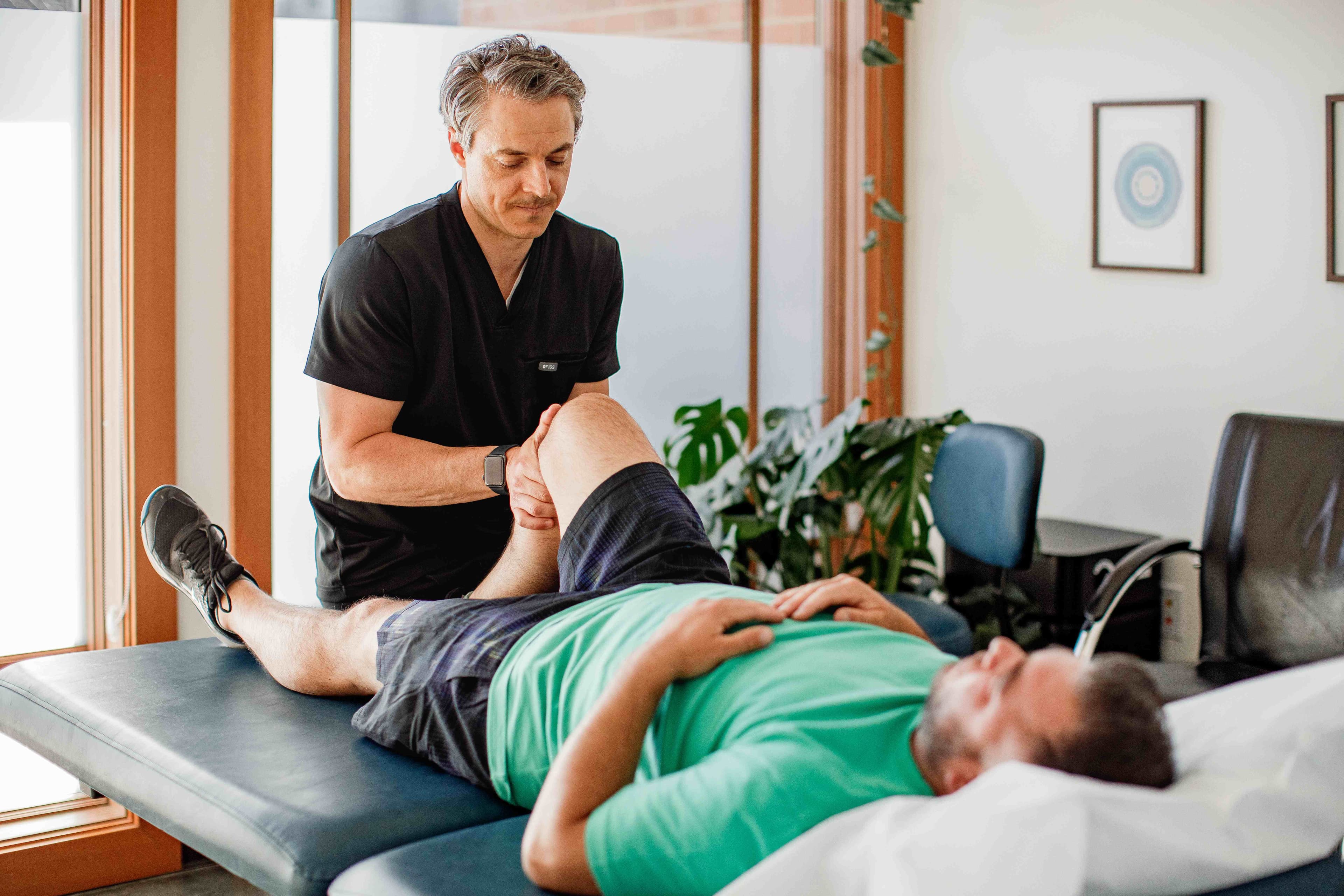
Sacroiliac joint pain treatment in Bellingham, WA
One of the most commonly missed sources of low back and buttock pain — with supportive evidence for carefully selected non-surgical treatments, including prolotherapy and PRP.
Book an initial visitA common cause of low back pain that imaging usually misses
Bottom line
SI joint dysfunction is responsible for a meaningful proportion of chronic low back and buttock pain — an estimated 15–30% of chronic low back pain cases. It is frequently underdiagnosed because standard MRI and X-ray are often normal. In the best available RCT, prolotherapy produced lasting pain relief in nearly 60% of patients at 15 months — versus 10% for corticosteroid.
The sacroiliac joints connect the sacrum to the iliac bones of the pelvis and transfer load between the spine and legs. Unlike most spinal joints, the SI joint has minimal inherent mobility — it is stabilized primarily by a dense network of posterior ligaments. When those ligaments are stressed, stretched, or incompletely healed, the joint becomes a reliable pain generator.
SI joint pain typically presents as one-sided low back pain below the belt line, often radiating into the buttock or posterior thigh. It is frequently worse with transitions — getting up from a chair, rolling over in bed, or stepping off a curb. It is a common sequela of pregnancy and childbirth, and a frequent contributor to back pain that persists despite normal imaging.
Cortisone into the SI joint often provides weeks of relief. Prolotherapy and PRP aim to address the ligament laxity and joint irritability that cortisone does not — and the RCT data show a clear and durable advantage at 15 months.
Diagnosis comes first
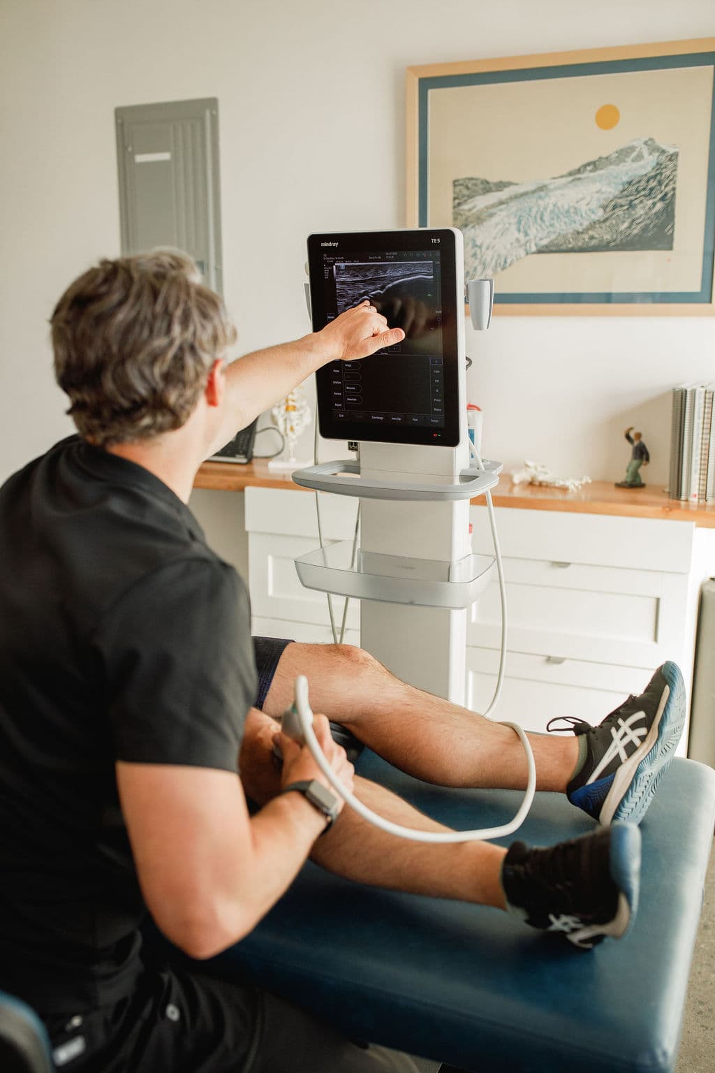
SI joint dysfunction is a clinical diagnosis. Dr. Borys uses a combination of provocation tests — FABER, Gaenslen, posterior shear, and thigh thrust — to assess whether the SI joint is the pain source before any treatment is considered. Ultrasound can confirm joint anatomy and guide injection placement; imaging is used to rule out structural causes rather than to diagnose SI joint pain, which is typically invisible on MRI or X-ray.
If the SI joint is not the primary pain source — or if there is a concurrent lumbar facet, disc, or piriformis component — Dr. Borys will identify this at the initial visit and address it accordingly.
Who comes to Dr. Borys with SI joint pain
- Have low back, buttock, or hip pain that has not resolved with rest, physical therapy, or cortisone
- Notice pain that worsens with transitions — standing up from sitting, rolling over in bed, or walking on uneven ground
- Have had a cortisone injection that helped briefly then faded
- Have been told imaging is normal but still have significant pain — SI joint dysfunction is typically invisible on MRI
What Dr. Borys uses for SI joint pain
Among regenerative options, prolotherapy has the strongest direct long-term RCT support for SI joint pain. PRP is used when a stronger biologic response is called for or when a shorter injection course is preferable. Shockwave addresses the soft-tissue component without injection.
Prolotherapy
Dextrose-based solution injected into the SI joint and posterior ligaments to stimulate a localized repair response. In the best available RCT (Kim 2010), prolotherapy produced ≥50% pain relief at 15 months in 58.7% of patients — versus 10.2% for corticosteroid. A short series of two to three injections is typical.
Learn more about prolotherapyPRP injections
Concentrated growth factors from your own blood placed into the SI joint or dorsal interosseous ligament. In one comparative study (Cusi et al.), PRP achieved similar outcomes to dextrose prolotherapy but required fewer injections to reach the same endpoint. Used when the clinical picture calls for a more potent biologic stimulus.
Learn more about PRP therapyShockwave therapy (ESWT)
Acoustic pressure waves applied to the posterior sacral ligaments and gluteal soft tissue. A non-injection option that can be used as a stand-alone treatment or in combination with prolotherapy or PRP when there is a significant soft-tissue component to the pain.
Learn more about shockwave therapyWhat the evidence shows
The SI joint evidence base is smaller than for knee OA or lateral epicondylitis. The foundational prolotherapy RCT includes a 15-month follow-up with a large and durable separation from steroid — which is uncommon in injection trial literature.
Prolotherapy vs corticosteroid for SI joint pain — 15-month RCT
Prospective randomized controlled trial (Kim et al., J Altern Complement Med 2010, n=48) comparing intra-articular dextrose prolotherapy to triamcinolone for confirmed SI joint pain. At 15 months, 58.7% of the prolotherapy group maintained ≥50% pain relief versus 10.2% in the steroid group (log-rank p < 0.005). Both groups improved equally at 2 weeks — the advantage of prolotherapy emerged over time and was sustained.
View on PubMedDextrose prolotherapy for musculoskeletal pain — systematic review
Systematic review (Hauser et al.) of dextrose prolotherapy across musculoskeletal conditions including ligament and joint pain. Found consistent evidence of pain reduction and functional improvement, supporting prolotherapy as an effective regenerative injection option for chronic joint pain.
View on PubMedPRP versus dextrose prolotherapy for SI joint pain — comparative study
Comparative study (Cusi et al.) of PRP versus dextrose prolotherapy for SI joint pain, finding that PRP achieved comparable outcomes to prolotherapy but required fewer injections to reach the same endpoint. Suggests PRP is an option for patients seeking a shorter treatment course.
View on PubMedPRP for SI joint dysfunction — systematic review and pooled analysis
Systematic review and pooled analysis (Goodwin et al., 2023) of PRP injection for sacroiliac joint dysfunction. Found clinically meaningful reductions in pain and functional improvement across included studies, with a favorable safety profile. The authors were careful to note the evidence base is still limited and does not yet establish PRP as superior to corticosteroid injection — small studies suggest it may offer sustained benefit for selected patients rather than a proven replacement for standard care.
View on PubMedInjective treatments for SI joint pain — systematic review and meta-analysis
Systematic review and meta-analysis (Ruffilli et al., 2024) of injective treatments for sacroiliac joint pain, including corticosteroid, PRP, and prolotherapy. Found evidence supporting regenerative injection options, particularly for patients who have not achieved lasting relief with corticosteroid alone.
View on PubMedWho is a reasonable candidate
Likely a good fit
- Have confirmed or clinically suspected SI joint dysfunction — pain reproduced by SI joint provocation tests
- Have chronic low back or buttock pain that has not responded to physical therapy or anti-inflammatories
- Have had temporary relief from a cortisone injection that faded within weeks to months
- Have posterior pelvic pain that worsens with single-leg loading, prolonged sitting, or stair climbing
- Are postpartum and have persistent pelvic girdle pain — a common and often underdiagnosed presentation
Less likely to benefit
- Have active infection, malignancy, or a bleeding disorder
- Are on anticoagulant therapy that cannot be safely held around the injection
- Have SI joint pain secondary to inflammatory arthropathy (ankylosing spondylitis, psoriatic arthritis) — these require rheumatologic management first
- Require surgical stabilization due to frank instability or structural failure — Dr. Borys will say so if this applies to you
Recovery timeline after treatment
Post-injection soreness
Increased aching in the low back, buttock, or posterior pelvis for a few days after injection is expected and is part of the intended healing response. Activity at a tolerable level is fine. Avoid NSAIDs — they blunt the repair process prolotherapy and PRP depend on.
Ice for short periods over the posterior pelvis can help if needed. Avoid anti-inflammatory medications for at least 5 days.
The quiet phase
Initial soreness settles but meaningful improvement may not yet be apparent. The joint and ligament environment is remodeling. Feeling close to your pre-injection baseline is normal and expected at this stage.
Check-in and possible repeat
Dr. Borys typically follows up at 6 weeks. Depending on the response, a second injection may be appropriate — the Kim RCT protocol used up to three injections, and most meaningful benefit builds over a series.
Continued improvement
The most durable improvements in the prolotherapy RCT data emerged beyond 3 months. Gradual, cumulative improvement is the typical pattern — not a single clear turning point.
Find out if your back pain is coming from the SI joint
The initial visit is a full evaluation — history, exam, review of any imaging. Dr. Borys evaluates whether the SI joint is a likely contributor to the pain and determines what next step, if any, fits the findings.
The first visit is an evaluation, not a commitment to any procedure.
Related conditions
Low back pain
Facet joint, ligament, and paraspinal pain — closely related to SI joint dysfunction and sometimes co-existing.
Prolotherapy
Dextrose-based regenerative injection targeting ligament laxity and joint stability — with the strongest long-term RCT support among regenerative options for SI joint pain.
Gluteal tendinopathy
Lateral hip tendon pain that can mimic or co-exist with SI joint dysfunction — worth distinguishing clinically.
Sacroiliac Joint Pain Treatment: Common Questions
How does prolotherapy work for SI joint pain?
A small volume of dextrose solution is injected into the SI joint and the posterior sacroiliac ligaments. The dextrose acts as a mild irritant that triggers a localized healing response — stimulating the production of collagen and connective tissue to reinforce the ligament laxity that is often the underlying driver of SI joint instability and pain. The effect builds over a series of two to three injections.
What is the difference between prolotherapy and PRP for the SI joint?
Both target the same structures — the SI joint and posterior ligaments. Dextrose prolotherapy is the more studied option, with a 15-month RCT showing durable superiority over corticosteroid. PRP delivers a more concentrated growth-factor stimulus and comparative evidence (Cusi et al.) suggests it may achieve similar results in fewer injections. The choice depends on clinical presentation, prior treatment history, and the degree of ligament involvement.
How is SI joint pain diagnosed?
SI joint dysfunction is a clinical diagnosis based on symptom pattern and physical examination. Dr. Borys uses a cluster of provocation tests — FABER, Gaenslen, posterior shear, and thigh thrust — to determine whether the SI joint is the primary pain source. Standard MRI and X-ray are typically normal with SI joint pain, which is why it is frequently missed. A positive response to a diagnostic block is the reference standard for confirming SI joint as the pain generator.
Can SI joint pain return after treatment?
The Kim RCT data showed 58.7% of patients maintained ≥50% pain relief at 15 months — meaning a meaningful proportion had durable benefit, but not all patients respond and not all responses are permanent. Contributing factors such as muscle weakness, movement patterns, and pelvic mechanics are addressed alongside injection treatment to support durability of results.
The information on this page is for general educational purposes only and is not individual medical advice. It is not a substitute for a consultation with a qualified provider. Whether a treatment is appropriate depends on your individual evaluation, and individual results vary.